
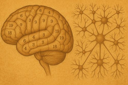
As neuroscience advances, the temptation will be to see each new map as definitive. Yet history counsels humility. Maps are tools, not truths.

By Matthew A. McIntosh
Public Historian
Brewminate
Introduction: The Brain as an Uncharted Landscape
When Korbinian Brodmann published his Vergleichende Lokalisationslehre der Großhirnrinde in 1909, he offered not just a classification of cortical regions but a way of seeing the brain as a territory to be mapped.1 His division of the cortex into numbered areas, based on cytoarchitectonic differences in cell structure, gave scientists a grid with which to navigate the enigmatic folds of the human cerebrum. It was a cartographic gesture, imposing borders and names where once there had been only undifferentiated terrain.
The history of brain mapping since Brodmann is not a straightforward tale of refinement but a shifting interplay of methods, metaphors, and technologies. From microscopic stains to functional imaging, from lesion studies to artificial intelligence, each stage reflects not only scientific advance but also the intellectual climate of its time. To follow this history is to watch science wrestle with the limits of analogy: the brain as map, as machine, as network.
Brodmann’s Areas and the Birth of Cortical Cartography
Cytoarchitecture and the Logic of Division
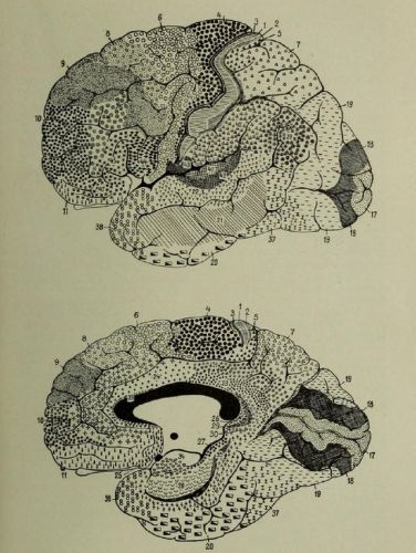
Brodmann’s project was rooted in the late nineteenth-century obsession with classification. Inspired by histological techniques developed by Franz Nissl, he sliced thin sections of brain tissue, stained them, and examined cellular arrangements. The differences he observed suggested that the cortex was not homogeneous but composed of distinct fields. His numbering system, still in use, provided a lingua franca for neuroscientists: Brodmann area 17 became synonymous with the primary visual cortex, area 44 with part of Broca’s language region.2
The appeal of Brodmann’s scheme lay in its apparent objectivity. Where phrenologists had once drawn speculative boundaries on the skull, Brodmann anchored his map in histology. Yet this objectivity was partial. His delineations were interpretive, reliant on his judgment about where one region ended and another began. The historian must note how readily the scientific community embraced his numbers as if they represented natural facts rather than human constructions.
The Authority and Limits of Brodmann’s Map
For decades, Brodmann’s atlas dominated neuroscience. It offered a stable scaffold upon which to build functional correlations. When Penfield stimulated cortical regions during neurosurgery, he often framed his findings with reference to Brodmann’s numbers. Yet the simplicity of the scheme also constrained inquiry. By treating the cortex as a mosaic of discrete units, Brodmann’s map obscured gradients, overlaps, and dynamic interactions. The tension between discrete localization and distributed function would haunt the next century of brain science.3
Mid-Twentieth Century: From Lesions to Electrophysiology
The Clinic as Laboratory
While Brodmann provided a morphological framework, the mid-twentieth century brought a wave of functional mapping grounded in pathology. Neurologists studied patients with localized brain damage, correlating deficits with anatomical lesions visible on postmortem dissection. This clinicopathological method, long practiced since Broca and Wernicke, gained new authority as imaging techniques improved. The brain became not just a stained specimen under the microscope but a site of clinical observation.
The logic here was inductive: if a stroke destroyed a region and language faltered, then that region must subserve language. Yet such reasoning risked reductionism. Lesions rarely respected neat boundaries. Compensation and plasticity blurred the clarity of causal inference. Still, these studies kept the mapping tradition alive, linking Brodmann’s histological areas to lived cognition.
Electrophysiology and the Live Brain
Meanwhile, electrophysiological methods opened another window. From Adrian’s recordings of neuronal firing to Penfield’s stimulation of awake surgical patients, the cortex could now be seen in action.4 Here mapping shifted from static grids to dynamic signals. The famous “homunculus” diagrams of motor and sensory cortices embodied this transition: the brain rendered not as histological patchwork but as a distorted map of the body itself.
This phase revealed a paradox. The more tools scientists gained, the more complex the brain appeared. Localization remained useful, but every new method undermined the notion of simple one-to-one correspondences.
The Age of Imaging: PET, fMRI, and Beyond
Visualizing Function in Real Time
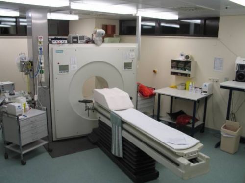
The late twentieth century ushered in a revolution. Positron emission tomography (PET) and functional magnetic resonance imaging (fMRI) allowed scientists to see living brains perform tasks in real time. Suddenly, cognition could be visualized as patterns of blood flow or metabolic activity.5 Maps proliferated, not of cytoarchitecture but of activation.
These methods carried seductive imagery. Brain scans, colored in reds and blues, became cultural icons, appearing in news articles and courtrooms. Yet historians must treat this imagery critically. The bright patches of an fMRI scan are not windows into thought itself but statistical inferences across many subjects. To map the brain this way is as much an act of construction as Brodmann’s stains.
Network Thinking and Connectivity
Imaging also shifted metaphors. Instead of discrete “centers,” scientists increasingly spoke of “networks.” The default mode network, the salience network, the executive network, all terms that frame the brain as a system of interacting nodes rather than isolated regions.6 Mapping now meant tracing connections, charting flows, drawing graphs. The cartographic impulse persisted, but the terrain had changed.
Twenty-First Century: Big Data, AI, and the Plastic Brain
The Human Connectome Project
In the twenty-first century, brain mapping has expanded into megascience. The Human Connectome Project, launched in 2009, sought to chart the entirety of neural connections using diffusion imaging and computational modeling.7 Where Brodmann had counted cells under a microscope, twenty-first-century scientists count connections by the billions. The scale is staggering, and so too are the metaphors: the brain as network, internet, or cosmos.
Artificial Intelligence as Cartographer
Artificial intelligence now enters the story not only as a subject but as a tool. Machine learning algorithms analyze neural data, detecting patterns no human eye could discern. In turn, AI models of neural networks draw inspiration from brain architecture, creating a recursive loop of influence. The map shapes the model, and the model reshapes the map.
Historians must ask what is gained and what is lost in this exchange. Just as Brodmann’s numbers imposed boundaries, AI’s pattern-recognition imposes its own logic, privileging what can be quantified and correlating at the risk of overinterpretation.
The Plastic Brain and Historical Reflection
Perhaps the greatest shift lies in the recognition of plasticity. Brodmann’s map implied fixed regions, stable borders. Modern neuroscience reveals constant change: neurons rewiring, networks reorganizing after injury, environments sculpting structure. The brain resists any final map. Each new cartography is provisional, contingent on method, metaphor, and moment.
Conclusion: Maps as Mirrors of Their Makers
The history of brain mapping from Brodmann to today is less a march of progress than a succession of lenses. Each generation drew its maps with the tools available and the metaphors it trusted: grids of numbered areas, distorted homunculi, glowing activation blobs, networks of nodes, and machine-detected patterns. None is wholly false, but none is final.
The historian sees here a broader truth. To map the brain is to mirror the culture that seeks to understand it. Brodmann’s grids reflected the classificatory zeal of early twentieth-century science. fMRI images reflect a visual culture enamored with color and immediacy. Network diagrams echo the digital age’s fascination with connectivity.
As neuroscience advances, the temptation will be to see each new map as definitive. Yet history counsels humility. Maps are tools, not truths. The brain, like the societies that study it, resists reduction. It is not a fixed territory but a living landscape, one that changes as much with the cartographer’s eye as with the subject itself.
Appendix
Footnotes
- Korbinian Brodmann, Vergleichende Lokalisationslehre der Großhirnrinde in ihren Prinzipien dargestellt auf Grund des Zellenbaues (Leipzig: Barth, 1909).
- Michael Petrides, et.al., “The Prefrontal Cortex: Comparative Architectonic Organization in the Human and the Macaque Monkey Brains,” Cortex 48:1 (2012), 50.
- Stanley Finger, Origins of Neuroscience: A History of Explorations into Brain Function (New York: Oxford University Press, 1994), 248–260.
- Wilder Penfield and Edwin Boldrey, “Somatic Motor and Sensory Representation in the Cerebral Cortex of Man as Studied by Electrical Stimulation,” Brain 60, no. 4 (1937): 389–443.
- Marcus E. Raichle, “Functional Brain Imaging and Human Brain Function,” Journal of Neuroscience 23, no. 10 (2003): 3959–3962.
- Olaf Sporns, “The Human Connectome: A Complex Network,” Annals of the New York Academy of Sciences 1224, no. 1 (2011): 109–125.
- David C. Van Essen et al., “The WU-Minn Human Connectome Project: An Overview,” NeuroImage 80 (2013): 62–79.
Bibliography
- Brodmann, Korbinian. Vergleichende Lokalisationslehre der Großhirnrinde in ihren Prinzipien dargestellt auf Grund des Zellenbaues. Leipzig: Barth, 1909.
- Finger, Stanley. Origins of Neuroscience: A History of Explorations into Brain Function. New York: Oxford University Press, 1994.
- Penfield, Wilder, and Edwin Boldrey. “Somatic Motor and Sensory Representation in the Cerebral Cortex of Man as Studied by Electrical Stimulation.” Brain 60, no. 4 (1937): 389–443.
- Petrides, Michael, et. al. “The Prefrontal Cortex: Comparative Architectonic Organization in the Human and the Macaque Monkey Brains.” Cortex 48:1 (2012), 46-57.
- Raichle, Marcus E. “Functional Brain Imaging and Human Brain Function.” Journal of Neuroscience 23, no. 10 (2003): 3959–3962.
- Sporns, Olaf. “The Human Connectome: A Complex Network.” Annals of the New York Academy of Sciences 1224, no. 1 (2011): 109–125.
- Van Essen, David C., et al. “The WU-Minn Human Connectome Project: An Overview.” NeuroImage 80 (2013): 62–79.
Originally published by Brewminate, 08.26.2025, under the terms of a Creative Commons Attribution-NonCommercial-NoDerivatives 4.0 International license.

